Ultrasound assessment
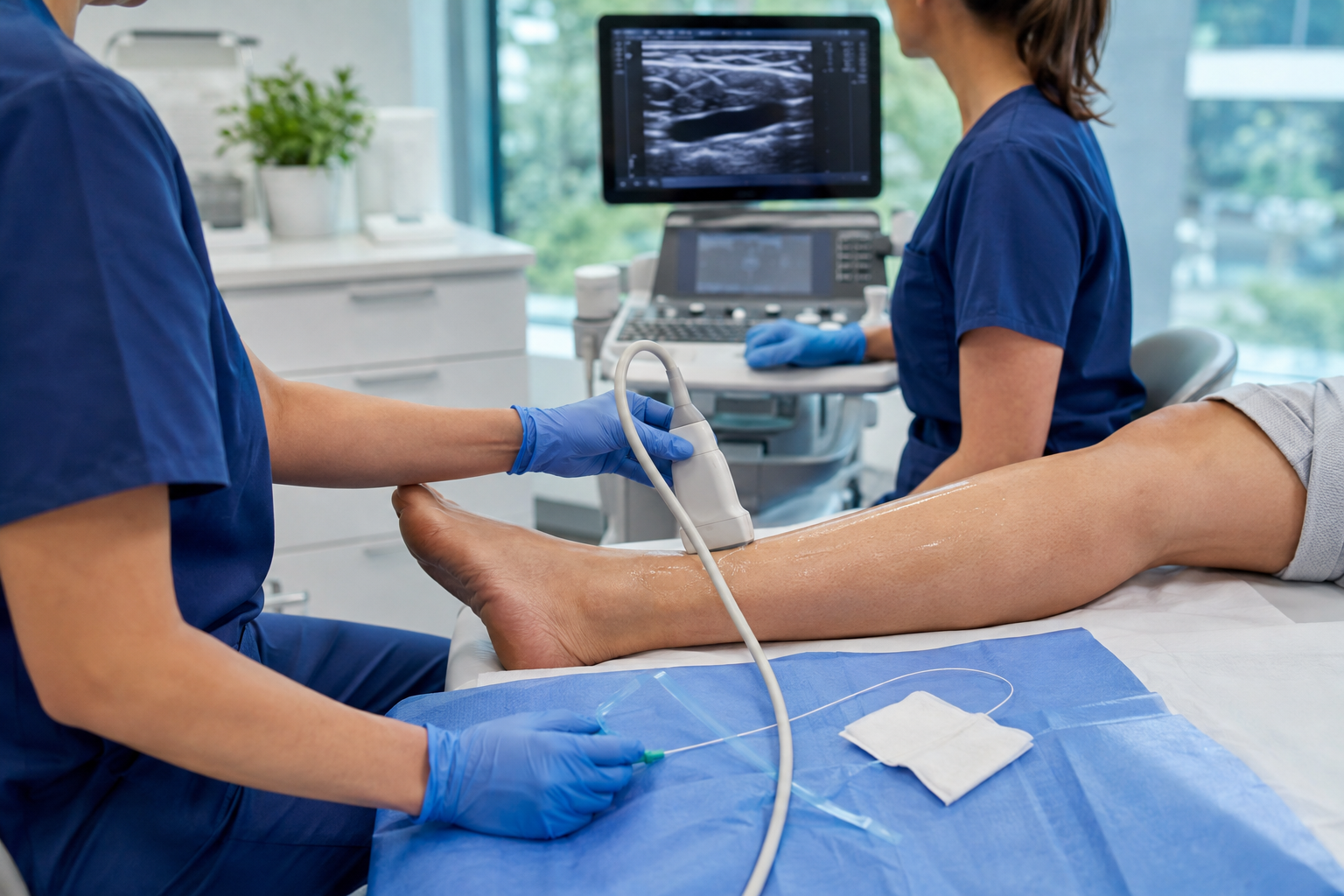
The target vein is assessed and mapped with duplex ultrasound. Access and treatment planning are determined by the treating clinician.
 Contact Medisol
Clinical education · Endovenous care
A practical overview of EVLA and EVRF workflows, technologies and treatment principles for healthcare professionals.
01 · EVLA
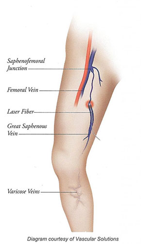
EVLA is a catheter-based, minimally invasive treatment used to close an incompetent superficial truncal vein, commonly the great or small saphenous vein. Duplex ultrasound is used to map reflux, plan access and guide the laser fibre to the intended treatment position. Current clinical guidance supports duplex-based treatment planning and recognises endothermal ablation as an established option for suitable patients with confirmed truncal reflux.
Laser energy is then delivered during a controlled, continuous withdrawal of the fibre. The resulting thermal effect contracts and closes the vein wall, redirecting venous return through competent pathways. Treatment planning and parameters remain the responsibility of the trained clinician and applicable product IFU.
How the technology works
EVLA combines image-guided fibre placement with perivenous tumescent anaesthesia and controlled thermal energy delivery. The treatment is continuous along the planned vein segment rather than delivered as separate segmental RF cycles.
The target vein is accessed percutaneously under ultrasound. An introducer is positioned and the laser fibre is advanced to the clinician’s planned starting point.
Tumescent solution is infiltrated around the vein to support local anaesthesia, compress the vein around the fibre and create separation from adjacent tissue.
Laser energy is applied as the fibre is withdrawn at a controlled rate. Wavelength, power, fibre design, pullback speed and vein anatomy all influence energy distribution.
Thermal interaction produces contraction and controlled injury of the vein wall. The treated segment occludes and subsequently undergoes fibrosis and gradual remodelling.
Typical clinical workflow
The target vein is assessed and mapped with duplex ultrasound. Access and treatment planning are determined by the treating clinician.
Under ultrasound guidance, the vein is accessed through a small puncture and an introducer is positioned.
A radial or bare laser fibre is advanced to the planned treatment position and checked before energy delivery.
Tumescent anaesthesia is delivered around the target vein to support patient comfort and separate the vein from surrounding tissue.
Laser energy is delivered while the fibre is withdrawn according to the clinician’s selected protocol, closing the treated vein.
The access site and treated vein are reviewed. Follow-up and aftercare are determined by the treating clinic.
Evidence snapshot · EVLA
NICE recommends endothermal ablation for suitable people with confirmed truncal reflux following duplex assessment.
SourceA 1470 nm systematic review reported pooled GSV occlusion of 93.9% at two years and 89.1% at five years across included studies.
SourceA review of 1920/1940 nm studies reported 96.3% pooled occlusion at 12 months, while noting protocol heterogeneity and limited long-term data.
SourcePublished outcomes are population-level findings and are not product-specific. Results vary with anatomy, technique, device, protocol, outcome definition and follow-up.
Potential clinical advantages
Related Medisol solutions
Explore laser systems, radial and bare fibres, introducer sets and vascular access products in the main product section.
Explore EVLA products02 · EVRF
EVRF is a minimally invasive thermal ablation technique in which a radiofrequency catheter is positioned inside an incompetent superficial vein under ultrasound guidance. Perivenous tumescence supports local anaesthesia, compresses the target vein and helps create a thermal buffer around the treatment segment.
Controlled energy heats the vein wall in defined treatment segments. The catheter is repositioned in measured increments until the planned length has been treated, producing contraction and closure of the target vein. Patient selection, access strategy, treatment parameters and surveillance are determined by the trained clinician and the applicable device instructions for use.
Educational procedure video
This manufacturer-provided clinical overview demonstrates catheter positioning, tumescent infiltration and controlled segment-by-segment radiofrequency energy delivery.
Watch on RF Medical’s YouTube channelTypical clinical workflow
The vein is mapped with ultrasound and accessed using an appropriate needle, guidewire and introducer.
The RF catheter is advanced through the introducer and positioned at the planned treatment point under ultrasound guidance.
Tumescent solution is infiltrated around the treatment segment and catheter position is reconfirmed.
The generator delivers radiofrequency energy using the selected temperature and cycle settings.
The catheter is withdrawn through the vein in controlled increments according to the device instructions and clinical protocol.
The catheter and introducer are removed, the access site is managed and the treated vein is reviewed by ultrasound.
Evidence snapshot · EVRF
A 2024 meta-analysis reported one-year GSV occlusion of 93.1% for RFA and 94.2% for laser ablation across the included evidence.
SourceThe same analysis found no statistically significant difference between RFA and laser for venous thrombotic events or phlebitis.
SourceMulti-society guidance supports thermal or non-thermal ablation according to anatomy, clinician expertise and informed patient preference.
SourcePublished outcomes are population-level findings and are not product-specific. Results vary with anatomy, technique, device, protocol, outcome definition and follow-up.
System and workflow benefits
Related Medisol solutions
Explore the endovenous RF platform, catheter options and vascular access products in the main product section.
Explore EVRF productsClinical context
EVLA and EVRF are established endovenous thermal techniques, but no single outcome applies to every patient, device or protocol. Duplex findings, anatomy, symptoms, comorbidities, treatment goals and the applicable IFU all inform clinical decisions.
Expected recovery considerations
Temporary tenderness, tightness, bruising and local inflammatory symptoms can occur following thermal ablation. Return to activity, compression and follow-up ultrasound should follow the treating clinic’s protocol and patient-specific assessment.
Recognised complications
Reported complications include superficial thrombophlebitis, pigmentation, sensory disturbance, skin burn, infection, thrombus extension or DVT, and later recanalisation or recurrent reflux. Incidence varies across technologies, techniques and study designs.
Selected clinical references
Discuss the workflow
3 Kirwan Place, Rouse Hill NSW 2155
Sydney, Australia